How to Keep Your Existing LDTs on the Market

How to Keep Your Existing LDTs on the Market

How to Keep Your Existing LDTs on the Market

Labs have some tough decisions to make now that the FDA has issued a final rule giving it authority to regulate LDTs. Labs offering LDTs prior to publication of the final rule on May 6 have three choices: 1) comply with the new FDA regs and keep their LDTs on inhouse test menus; 2) switch to an FDA-cleared test kit (if available); or 3) ignore the FDA regs and risk potential enforcement actions and fines. For expert advice on option 1 from regulatory attorney Christine Bump.

Christine Bump, a regulatory attorney at Penn Avenue Law & Policy (Washington, DC), has been guiding laboratories and test manufacturers through the FDA’s premarket clearance process and post market compliance requirements for 20 years. Below we summarize Bump’s
advice for keeping existing LDTs on the market.

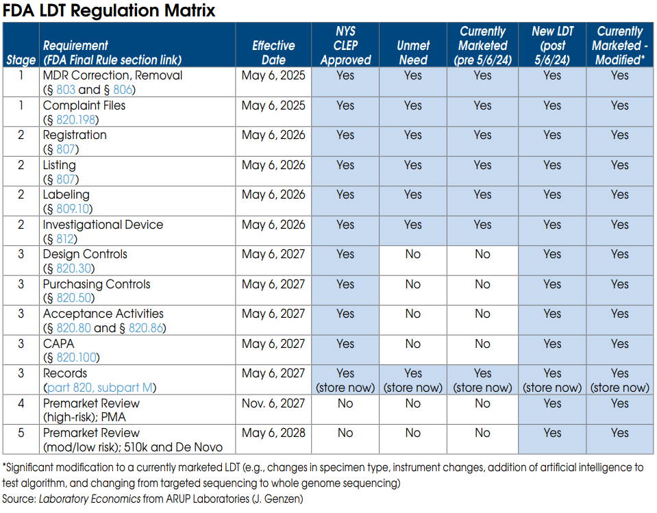
FDA Stage 1 requirements for currently marketed LDTs
Nearly all LDTs, including currently marketed LDTs (prior to May 6, 2024), “unmet need” LDTs performed by “integrated” health systems and NYS CLEP-approved tests, must meet Stage 1 requirements by May 6, 2025.

Stage 1 includes FDA Medical Device Reporting (MDR), which will require laboratories to report adverse events for any LDT that they perform. Under FDA’s regulations, an adverse event is any event that reasonably suggests that a device has or may have caused or contributed to a death or serious injury, or would be likely to cause or contribute to a death or serious injury if the event happens again. An adverse event report would therefore be required for an incorrect test result that has, may, or could cause such consequences. The incorrect test result could be caused by instrument malfunctions as well as mislabeled or contaminated specimens.

An adverse event report must generally be reported to the FDA within 30 days after the lab became aware of the error. The regulations require specific information be included in the report, and labs will also need to file\ a report that describes what corrective action they took, or if they chose to remove that LDT from their test menu.

An adverse event report could prompt the FDA to ask follow-up questions or schedule an on-site inspection.

The FDA is especially sensitive to incorrect test results that delayed patient treatment or caused
unnecessary treatment (or had the potential to do so).

Stage 1 also requires labs to maintain complaint files for each LDT they offer, including the date the complaint was received; the name, address, and phone number of the complainant; the nature and details of the complaint; any corrective action taken, etc. Specific records and reports
must also be maintained and submitted regarding corrections and removals of tests, including for
repairs, adjustments, relabeling, etc.

FDA Stage 2 requirements for currently marketed LDTs
Once again, Stage 2 requirements apply to nearly all LDTs and become effective May 6, 2026.

Stage 2 requires each laboratory to be registered with the FDA and list all of the LDTs they perform.

Stage 2 also requires labs to submit labeling for each LDT they offer. Labeling includes test performance information and a summary of supporting validation. As part of its review of labeling, the FDA plans to look closely at claims of superior performance and whether those claims are adequately substantiated. This includes any test claims made on a lab’s website, brochures or by sales reps.

Labeling information is typically included as a package insert or affixed to test kit box for FDA-cleared or approved IVD tests. However, since LDTs are not distributed in boxed test kits, it is unclear exactly where the label for an LDT will need to be placed. Labels might be required on the LDT test requisition form, but this hasn’t been confirmed yet. We’re waiting for the FDA to issue more guidance on label requirements.

FDA Stage 3 requirements for currently marketed LDTs

By May 6, 2027, currently marketed as well as “unmet need” LDTs developed and used within “integrated” health systems need to be in compliance with the records requirements of the quality system regulation. These records relate to the device master record, the device history record, and the quality system record. (Note: Complaint file record requirements are already covered under Stage 1.)

The device master records must include information about device specifications, production process specifications, quality assurance procedures and specifications, packaging and labeling specifications, and procedures and methods for installation, maintenance, and servicing.

The device history records must include information about the dates and quantity manufactured, the quantity released for distribution, acceptance records, information to identify each production unit and device identifiers.

Labs must have the required information compiled, documented, and in a records system that is accessible and readily available to FDA investigators during inspections.

FDA On-site Inspections
All registered lab facilities performing LDTs are subject to scheduled on-site inspections by the FDA every two years. FDA inspections are completely different than and are independent of CMS CLIA and CAP inspections. FDA inspectors will be focused on reviewing LDT test records and files. However, the reality is that the FDA already lacks the resources to perform regularly scheduled on-site inspections of existing test kit manufacturers. The agency may have difficulty keeping up with the thousands of new lab facilities that fall under its purview as a result of its final rule. 
Google Releases AI Tool for Pathology

Google Releases AI Tool for Pathology

Google Releases AI Tool for Pathology

Google has released a free cloud-based tool (“Path Foundation”) that 
transforms digitized slide images into numerical data that researchers 
can use to create AI tools for pathology. “This is a landmark for us,” says 
David Steiner, MD, PhD, Clinical Research Scientist at Google. Based on  feedback to Path Foundation, Google will make decisions toward  developing commercial AI tools for pathologists. 

For more than seven years, Google has employed a team of clinical research scientists based in Mountain View, California and London, England, tasked with developing AI software tools for radiology and pathology. Below is a summary of LE’s interview with Google’s David Steiner, MD, PhD, Clinical Research Scientist.

How can labs and pathologists use Path Foundation?

Commercial labs and academic medical centers, for example, can use Path Foundation to convert patches of their digitized slide images into numerical vectors, known as embeddings. These embeddings capture important features and patterns contained in digitized slide images which are learned by Path Foundation during its training on millions of pathology images. After the embeddings for each image are collected, this data can be used to train and create custom algorithms for a range of tasks such as identifying tissue type, tumors, or performing quality assurance on digitized images.

What’s the benefit of using a “Foundation” model to analyze content in images?
Leveraging the embeddings from Path Foundation requires less data and computational resources than traditional methods, giving researchers a big head start toward developing their own algorithms.

In contrast, traditional methods such as strongly supervised deep learning models require more resources because every task requires fresh training in its own unique deep learning model and many labeled images for each category of interest.

The foundation model is similar to that of seasoned guitar player quickly learning a new song by ear. Because the guitar player has already built up a foundation of skill and understanding, they can quickly pick up the patterns and groove of a new song.

Where did you get the digitized images to train Path Foundation on?
We used 20,000 whole-slide images, covering millions of image patches, from a variety of sources, including the National Cancer Institute’s Cancer Genome Atlas, as well as academic medical centers and some private pathology labs.

What is the user roadmap to getting started with Path Foundation?
After filling out the access form, users can run a small demo notebook that walks them through how to train a tumor classifier. To use Path Foundation on their own task (i.e., identifying tissue type) they would need to collate a set of digital pathology images with accompanying labels. The user would then upload these images and labels into Google Cloud.

From there, the users can adapt the demo notebook to call the Path Foundation API on their uploaded images. Using the existing code in the demo notebook they would then train a model to classify their images based on the labels and evaluate its performance on a held-out part of
the dataset not used to train the model. In the future we hope to make it even easier to use Path Foundation and the embeddings directly in your pathology slide viewer with no coding required.

We’re offering Path Foundation free to users on github.com/Google-Health.

Do labs that use Path Foundation need to share their digitized images or the results of their research with Google?
They don’t need to share the images or the results of their research with Google. They do need to have their images stored in Google Cloud (in their own private or institution account), but these are kept private to the user and not accessible by Google. In addition, when the images are sent to the Path Foundation model to compute the embeddings, they are not saved or stored by Google.

Any plans to develop commercial AI software tools like Ibex, Paige or PathAI?
Yes. Path Foundation represents a landmark toward that end.

What is the next step for your research team in terms of developing AI tools for pathologists?
We’ll take feedback on users from Path Foundation to understand key-use cases and how to make the tool better and easier to use. We’ll also explore how these embeddings might be used with large language and large multimodal models (LLMs & LMMs). And then we plan to develop useful approaches and models for working with whole slide images (WSIs), in addition to the “patch-based” models and applications that this current tool represents.

Has Google completed any studies related to AI-assisted diagnostic tools for cancer?
Yes. We have published several studies in peer-reviewed journals.

Most recently, we published a study that used AI to predict immunotherapy outcomes from digitized slide images in non-small cell lung cancer (Cancer Research, vol. 84, 2024).

In 2023, we published a paper that showed how AI can be used for clinical decision-making in colorectal cancer (Nature Communications Medicine, vol. 3, 2023). And, in 2022, we published a study that used AI for diagnosis and Gleason Grading of prostate cancer (Nature Medicine, vol. 28, 2022).

We have also published studies focusing on AI models for breast cancer.

Will AI algorithms eventually replace pathologists?
AI will make pathologists better rather than replace them. Initially, AI will be used to automate repetitive tasks such as locating the image patches that pathologists should focus their eyes on. Eventually, AI could be used to query images. Pathologists and researchers may someday be able to
type in specific questions and get AI answers about an image.

Is there the potential to integrate pathology and radiology image data using AI?
I’m excited to explore this and do see the promise in bringing these two specialties together. I believe the combination will yield more than the sum of the parts. Google does have a team of research scientists working on AI tools for radiology. And we did introduce an AI tool for chest x-rays (CXR Foundation) in July 2022.

Pathologist Job Openings Remain Near Record-High

Pathologist Job Openings Remain Near Record-High

Pathologist Job Openings Remain Near Record-High

The biggest online job board for pathologists, PathologyOutlines.com, currently has 705 pathologist jobs listed. That’s near the site’s all-time record of 706 pathologist job ads reached in February 2022. Prior to 2020, the average number of job openings was between 300 and 400.

Rich Cornell, President and Founder of the life sciences recruiting firm Santé Consulting (St. Louis, MO), says it’s important for labs to understand that the shortage is not going away soon. Cornell says, “Labs must establish an internal sense of urgency in the hiring process in order to hire effectively in this market. The total actual number of U.S. pathologist openings is currently closer to 1,000 when including jobs that are not advertised on PathologyOutlines.com. The biggest competition exists for jobs in the subspecialties in highest demand: cytopathology, hematopathology and gastrointestinal.

According to Cornell, only about 600 pathologist residents and fellows graduate each year. A large percentage of those graduates will require visa sponsorship, but only about 1/3 of visa requests will likely get accepted. In other words, the market is out of balance and will continue this way for the next several years, says Cornell.

So what should labs do in this tight job market? Cornell says it boils down to these two things:

1. The interview-to-hire ratio has changed. Pathology practices should expect to interview an average of five candidates in order to successfully hire one pathologist. That’s the new average.

2. Young millennial pathologists (age 27-42 years) are seeking low stress working environments and a quick interview timeframe. They expect offers within 48 hours after an interview. The timng and pace of your offer are crucial once the interview takes place.

How are other labs advertising for pathologist openings in light of this severe shortage? Here are some recent examples from PathologyOutlines.com:

$100K Sign-On Bonus
A large independent pathology practice in Connecticut is offering $100,000 sign-on bonuses in an effort to hire one or more full-time surgical pathologists with subspecialty training in gastrointestinal pathology, breast or hematopathology. The annual salary range is highly competitive with four weeks vacation. Current fellows, pathologists straight out of fellowships and experienced pathologists are encouraged to apply.

22 Pathologists Needed at HCA Healthcare
HCA Healthcare has ads for 22 pathologist job openings—mostly for its hospitals in Florida, Georgia and Texas. HCA Healthcare is offering sign-on bonuses of up to $30,000. Other organizations with a large number of job ads include Sonic Healthcare (21 pathologist openings), Rutgers New Jersey Medical School (11 openings) and Northwell Health in New York (10 openings).

Cornell says that many groups are struggling because of their hiring process, or lack thereof. “We recently worked with a large healthcare system with more than 20 pathologists. They needed four additional pathologists to handle their workload volume increases. However, their internal process prevented them from being successful. They would take 4 weeks to make an offer once the interview had occurred. Candidates would lose interest because they were left waiting for too long after they were interviewed. Younger candidates expect feedback from employers immediately following the interview.”

PathAI Announces 13 Lab Contracts for Its AI Tools

PathAI Announces 13 Lab Contracts for Its AI Tools

PathAI Announces 13 Lab Contracts for Its AI Tools

ePathAI (Boston, MA) has won contracts with 13 lab organizations that will be using its FDA-cleared AISight digital pathology image management and viewer analysis system. These new clients will also be using PathAI’s software algorithm AIM-PD-L1 NSCLC RUO, which quantitates the percentage of PD-L1 positive tumor and immune cells in non-small cell lung cancer samples.

Among the 13 labs that will be using PathAI’s AI-based software algorithms is PathAI Diagnostics (Memphis, TN)—formerly named Poplar Healthcare. PathAI acquired Poplar Healthcare in August 2021 (see LE, August 2021). PathAI Diagnostics is a full-service anatomic pathology lab with 350 employees, including 25 pathologists.

Other labs that will be using PathAI’s software tools include Caris Life Sciences, NeoGenomics and University of Pittsburgh Medical Center (see table below).

These labs will have early access to additional AI algorithms that PathAI expects to bring to market for research use only (RUO) within the next few months. These products include AI software tools for quantitative PD-L1 tests for melanoma, head and neck, and bladder cancer, as well as an
algorithm for the automated quantification of HER2 IHC images in breast cancer tissue.

Labs will have the potential to validate PathAI’s RUO products so that they can be used as lab-developed tests for clinical diagnostics, according to Andy Beck, MD, PhD, Chief Executive of PathAI.

The new clients were all signed by PathAI within the past six months. Beck anticipates more lab client announcements soon. Eric Walk, MD, Chief Medical Officer, and Parth Chodavadia, Head of Commercial, Digital Diagnostics, are leading the marketing efforts at PathAI.

PathAI has raised a total of $255 million since being formed in 2016. Major backers include Labcorp, Kaiser Permanente, Merck and Bristol Meyers, as well as the private equity firms General Atlantic and General Catalyst.

Spotlight Interview with Proscia’s Nathan Buchbinder

Spotlight Interview with Proscia’s Nathan Buchbinder

Spotlight Interview with Proscia’s Nathan Buchbinder

Proscia Inc. (Philadelphia, PA) markets a digital pathology software platform (Concentriq) that helps upload, organize into patient cases, annotate, and store whole slide images. Concentriq is currently being used by more than 6,000 scientists and pathologists at 300+ clinical and research organizations around the world. Proscia has also developed AI-based applications, including a program for quality control of digitized images (currently for the research market only). Here’s a summary of our recent interview with Nathan Buchbinder, Co-Founder & Chief Product Officer at Proscia.

Describe when and who founded Proscia.
Proscia was founded in 2014 by myself and two other computer scientists from Johns Hopkins University and University of Pittsburgh. These include our Chief Executive David West and our Chief Technology Officer Coleman Stavish. We currently have 100 employees.

How much capital has Proscia raised?
We raised $37 million in June 2022 bringing our total funding to $72 million. More than 10 private equity firms have invested in Proscia, including the following that have board seats: Emerald Development Managers, Flybridge Capital Partners, Razor’s Edge Ventures and Scale Venture Partners.

Why were drug development research firms so quick to adopt digital pathology?

Because their return on investment (ROI) on digitizing slides was self-evident and nearly immediate. Ten of the top 20 pharmaceutical companies, including Amgen, Bayer and Bristol Myers Squibb, are using Proscia to help manage their digitized slide images. These companies operate research sites and collaborate with third-party contract research organizations all around the world. Concentriq gives them a single hub where digitized slide images can be accessed and shared.

What’s the current status of digital pathology for clinical diagnostics?
Adoption started much slower in the clinical market. Some of our early adopters, including Thomas Jefferson University Hospitals and Johns Hopkins’ Department of Pathology, initially used digital pathology primarily for research and education.

However, over the past two years, we’ve seen a huge surge in demand from the clinical market, including integrated delivery networks, reference labs and even smaller pathology practices (~5 pathologists). These labs are using digital pathology for peer reviews, conferencing, consults, and tumor boards, as well as primary diagnosis of cancer cases. Most of our customers are in life sciences and research, but that’s quickly changing.

What’s your advice for pathology labs planning to transition to digital pathology?
Number one, get everyone involved at the start, not just executives and pathologists, but lab managers and histotechs. Number two, don’t underestimate the value of having an archive of digitized slides, not only in terms of internal research and education, but also its value to third-
party life sciences and pharmaceutical companies.

What’s your outlook for digital pathology adoption in the United States?
It will be widespread with nearly 100% adoption within five years. Drivers include the new Category III CPT codes for digital pathology and the potential for Medicare reimbursement. In addition, the application of AI, which requires digitized slides, will increase pathologist accuracy and efficiency.

The shift from microscope to monitor will be transformational. Winners and losers will be determined based on how fast and how well they implement technology. It could help the biggest commercial labs gain share in anatomic pathology or result in a different outcome that we can’t imagine today.

Did Digital Pathology Utilization Increase During The Pandemic?

Did Digital Pathology Utilization Increase During The Pandemic?

Did Digital Pathology Utilization Increase During The Pandemic?

The conventional wisdom says that digital pathology use surged as a result of the pandemic. However, Medicare data for CPT 88361 (computer-assisted IHC for breast cancer) tells a different story. The volume of Medicare Part B allowed claims for 88361 declined by 16% to 160,819 in 2020, followed by only a 3% rebound to 165,231 in 2021. CPT 88361 is the only code devoted specifically to bill Medicare for reading digitized slides. It therefore gives an indication of digital pathology trends in the clinical market.

Another indication that the digital pathology market has not taken off during the pandemic is the falling number of pathologists using it. A total of 736 pathologists billed Medicare for CPT 88361 in 2020 (the latest year of available data), which was down from 871 pathologists in 2019. The number of independent labs billing Medicare for CPT 88361 declined from 96 labs in 2013 to 66 labs in 2019 but increased slightly to 69 labs in 2020.

The main barrier, irrespective of the pandemic, to more widespread adoption of digital pathology has been the added expense of digitizing slides without reimbursement. The problem is that digital pathology comes as an “add on” process that is produced from a traditional glass slide. Digital pathology does not eliminate the need to process, section, glass-slide-mount and stain biopsy specimens. A high-end conventional microscope costs between $9,000 and $12,000, while a complete digital pathology
system can cost between $100,000 and $400,000.

In addition, pathologist practice patterns are hard to change, especially without a clear clinical benefit and/or compelling financial incentive.

 Artificial intelligence could be the game changer that jumpstarts the digital pathology market. AI-based decision-support tools that boost pathologist productivity and reduce errors need digitized images to read. AI vendors (PathAI, Paige, Ibex Medical Analytics, etc.) claim their software can help pathologists read 30+% more slides per day. This may provide hospitals and labs with the return on investment necessary to justify an investment in digital pathology scanners.